


 Foundational characteristics of cancer include proliferation, angiogenesis, migration, evasion of apoptosis, and cellular immortality. Find key markers for these cellular processes and antibodies to detect them.
 The SUMOplot™ Analysis Program predicts and scores sumoylation sites in your protein. SUMOylation is a post-translational modification involved in various cellular processes, such as nuclear-cytosolic transport, transcriptional regulation, apoptosis, protein stability, response to stress, and progression through the cell cycle.
 The Autophagy Receptor Motif Plotter predicts and scores autophagy receptor binding sites in your protein. Identifying proteins connected to this pathway is critical to understanding the role of autophagy in physiological as well as pathological processes such as development, differentiation, neurodegenerative diseases, stress, infection, and cancer.
Beta-Actin Antibody
Mouse Monoclonal Antibody (Mab)
- SPECIFICATION
- CITATIONS
- PROTOCOLS
- BACKGROUND

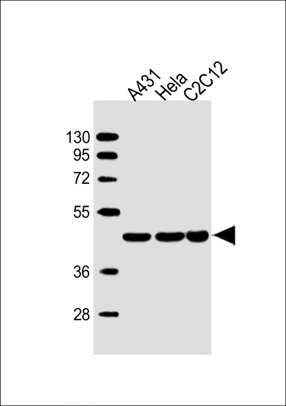
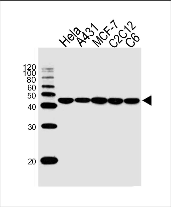
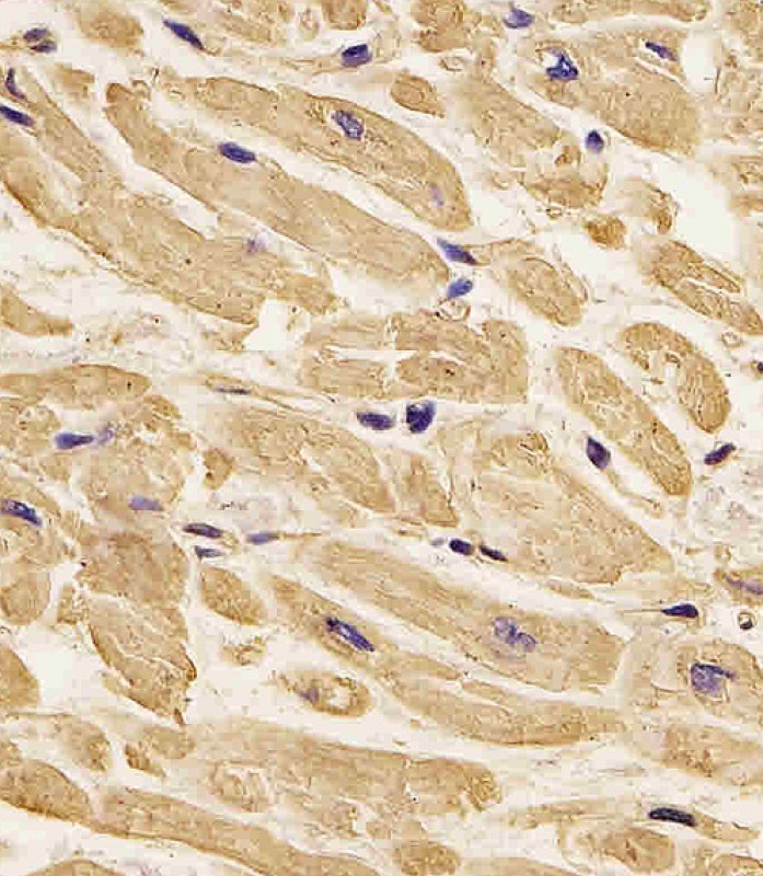
Application
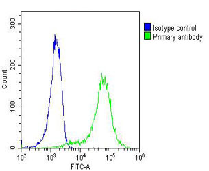
| WB, IHC, FC |
|---|---|
| Primary Accession | P60709 |
| Other Accession | A2BDB0, P63259, P63260, P63261, Q5ZMQ2, P63258, P60711, Q6QAQ1, P60710, Q4R561, P60706, P60712, P53505, P60708, P60713 |
| Reactivity | Human, Rat |
| Predicted | Xenopus, Bovine, Chicken, Horse, Monkey, Mouse, Pig, Sheep |
| Host | Mouse |
| Clonality | Monoclonal |
| Calculated MW | H=42;M=42;Rat=42 KDa |
| Isotype | IgG1,Igk |
| Antigen Source | HUMAN |
| Gene ID | 60 |
|---|---|
| Antigen Region | 11-395 aa |
| Other Names | ACTB; Actin, cytoplasmic 1; Beta-actin; Actin, cytoplasmic 1, N-terminally processed |
| Dilution | WB~~1:1000 IHC~~1:25 FC~~1:25 |
| Target/Specificity | This ACTB Monoclonal antibody is generated from mouse immunized with ACTB recombinant protein. |
| Format | Purified monoclonal antibody supplied in PBS with 0.09% (W/V) sodium azide. This antibody is purified through a protein G column, followed by dialysis against PBS. |
| Storage | Maintain refrigerated at 2-8°C for up to 2 weeks. For long term storage store at -20°C in small aliquots to prevent freeze-thaw cycles. |
| Precautions | Beta-Actin Antibody is for research use only and not for use in diagnostic or therapeutic procedures. |
| Name | ACTB |
|---|---|
| Function | Actin is a highly conserved protein that polymerizes to produce filaments that form cross-linked networks in the cytoplasm of cells (PubMed:29581253). Actin exists in both monomeric (G-actin) and polymeric (F-actin) forms, both forms playing key functions, such as cell motility and contraction (PubMed:29581253). In addition to their role in the cytoplasmic cytoskeleton, G- and F-actin also localize in the nucleus, and regulate gene transcription and motility and repair of damaged DNA (PubMed:29925947). Part of the ACTR1A/ACTB filament around which the dynactin complex is built. The dynactin multiprotein complex activates the molecular motor dynein for ultra-processive transport along microtubules (By similarity). |
| Cellular Location | Cytoplasm, cytoskeleton. Nucleus Note=Localized in cytoplasmic mRNP granules containing untranslated mRNAs. |

Thousands of laboratories across the world have published research that depended on the performance of antibodies from Abcepta to advance their research. Check out links to articles that cite our products in major peer-reviewed journals, organized by research category.
info@abcepta.com, and receive a free "I Love Antibodies" mug.

Provided below are standard protocols that you may find useful for product applications.
Background
This gene encodes one of six different actin proteins. Actins are highly conserved proteins that are involved in cell motility, structure, and integrity. This actin is a major constituent of the contractile apparatus and one of the two nonmuscle cytoskeletal actins.
References
Sex-specific proteome differences in the anterior cingulate cortex of schizophrenia. Martins-de-Souza D, et al. J Psychiatr Res, 2010 Apr 8. PMID 20381070. Identification of a hormone-regulated dynamic nuclear actin network associated with estrogen receptor alpha in human breast cancer cell nuclei. Ambrosino C, et al. Mol Cell Proteomics, 2010 Jun. PMID 20308691. Contribution of rearranged actin structures to the spread of Ectromelia virus infection in vitro. Boratynska A, et al. Acta Virol, 2010. PMID 20201613. Molecular mechanisms underlying nucleocytoplasmic shuttling of actinin-4. Kumeta M, et al. J Cell Sci, 2010 Apr 1. PMID 20197409. Tyrosine phosphorylation of cofilin at Y68 by v-Src leads to its degradation through ubiquitin-proteasome pathway. Yoo Y, et al. Oncogene, 2010 Jan 14. PMID 19802004.
If you have used an Abcepta product and would like to share how it has performed, please click on the "Submit Review" button and provide the requested information. Our staff will examine and post your review and contact you if needed.
If you have any additional inquiries please email technical services at tech@abcepta.com.
Ordering Information
Other Products
Shipping Information