


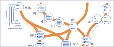


 Foundational characteristics of cancer include proliferation, angiogenesis, migration, evasion of apoptosis, and cellular immortality. Find key markers for these cellular processes and antibodies to detect them.
 The SUMOplot™ Analysis Program predicts and scores sumoylation sites in your protein. SUMOylation is a post-translational modification involved in various cellular processes, such as nuclear-cytosolic transport, transcriptional regulation, apoptosis, protein stability, response to stress, and progression through the cell cycle.
 The Autophagy Receptor Motif Plotter predicts and scores autophagy receptor binding sites in your protein. Identifying proteins connected to this pathway is critical to understanding the role of autophagy in physiological as well as pathological processes such as development, differentiation, neurodegenerative diseases, stress, infection, and cancer.
Phospho-Bad(S99) Antibody
Affinity Purified Rabbit Polyclonal Antibody (Pab)
- SPECIFICATION
- CITATIONS
- PROTOCOLS
- BACKGROUND

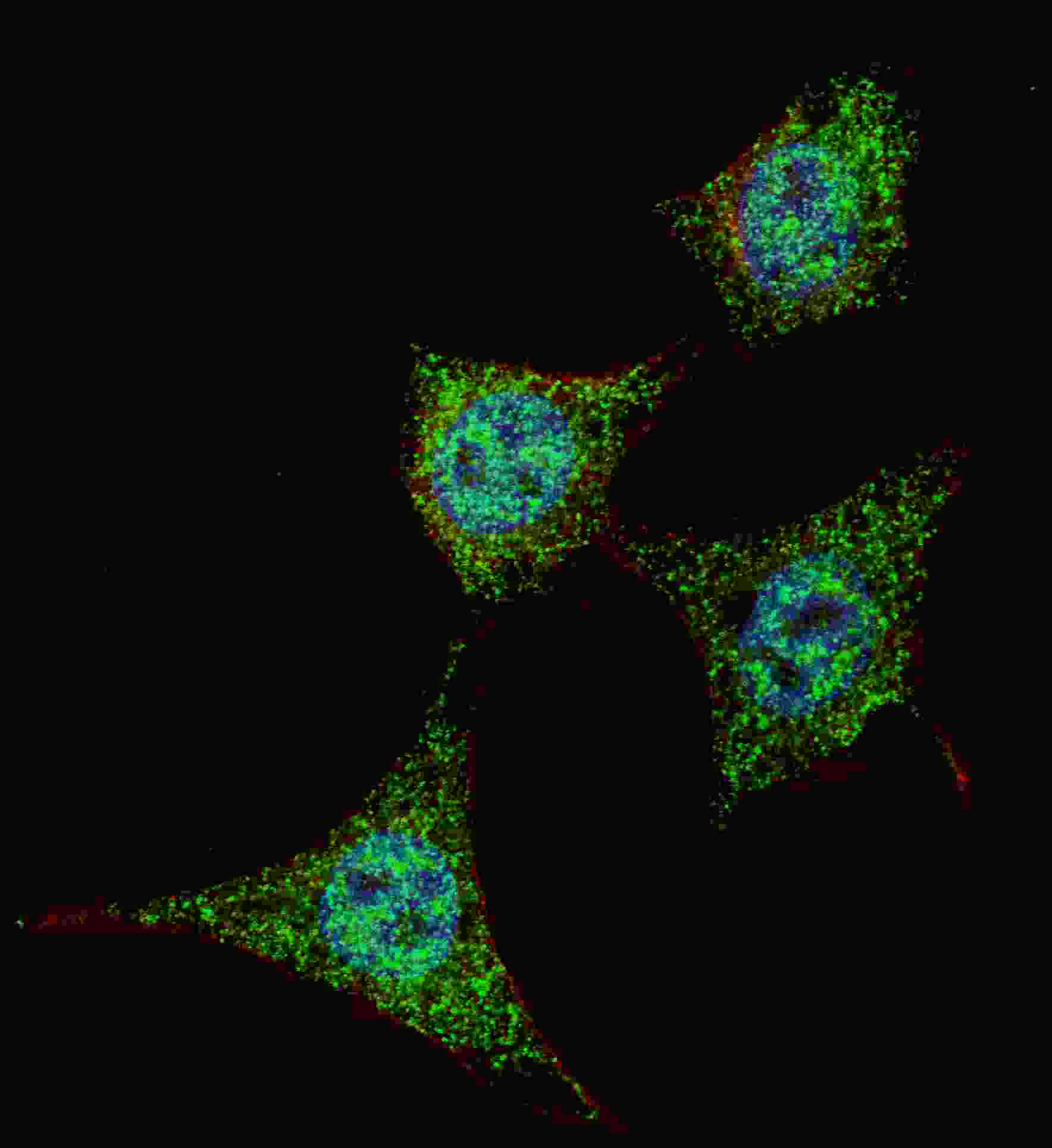
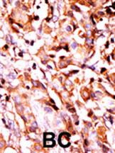
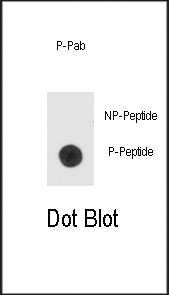
Application
| IHC-P, DB, IF, E |
|---|---|
| Primary Accession | Q92934 |
| Other Accession | O35147, Q61337 |
| Reactivity | Human |
| Predicted | Mouse, Rat |
| Host | Rabbit |
| Clonality | Polyclonal |
| Isotype | Rabbit IgG |
| Calculated MW | 18392 Da |
| Gene ID | 572 |
|---|---|
| Other Names | Bcl2-associated agonist of cell death, BAD, Bcl-2-binding component 6, Bcl-2-like protein 8, Bcl2-L-8, Bcl-xL/Bcl-2-associated death promoter, Bcl2 antagonist of cell death, BAD, BBC6, BCL2L8 |
| Target/Specificity | This Bad Antibody is generated from rabbits immunized with a KLH conjugated synthetic phosphopeptide corresponding to amino acid residues surrounding S99 of human Bad. |
| Dilution | IF~~1:200 IHC-P~~1:50~100 DB~~1:500 |
| Format | Purified polyclonal antibody supplied in PBS with 0.09% (W/V) sodium azide. This antibody is purified through a protein A column, followed by peptide affinity purification. |
| Storage | Maintain refrigerated at 2-8°C for up to 2 weeks. For long term storage store at -20°C in small aliquots to prevent freeze-thaw cycles. |
| Precautions | Phospho-Bad(S99) Antibody is for research use only and not for use in diagnostic or therapeutic procedures. |
| Name | BAD |
|---|---|
| Synonyms | BBC6, BCL2L8 |
| Function | Promotes cell death. Successfully competes for the binding to Bcl-X(L), Bcl-2 and Bcl-W, thereby affecting the level of heterodimerization of these proteins with BAX. Can reverse the death repressor activity of Bcl-X(L), but not that of Bcl-2 (By similarity). Appears to act as a link between growth factor receptor signaling and the apoptotic pathways. |
| Cellular Location | Mitochondrion outer membrane. Cytoplasm {ECO:0000250|UniProtKB:Q61337}. Note=Colocalizes with HIF3A in the cytoplasm (By similarity). Upon phosphorylation, locates to the cytoplasm. {ECO:0000250|UniProtKB:Q61337} |
| Tissue Location | Expressed in a wide variety of tissues. |

Thousands of laboratories across the world have published research that depended on the performance of antibodies from Abcepta to advance their research. Check out links to articles that cite our products in major peer-reviewed journals, organized by research category.
info@abcepta.com, and receive a free "I Love Antibodies" mug.

Provided below are standard protocols that you may find useful for product applications.
Background
Bad is a member of the BCL-2 family. BCL-2 family members are known to be regulators of programmed cell death. This protein positively regulates cell apoptosis by forming heterodimers with BCL-xL and BCL-2, and reversing their death repressor activity. Proapoptotic activity of this protein is regulated through its phosphorylation. Protein kinases AKT and MAP kinase, as well as protein phosphatase calcineurin are found to be involved in the regulation of this protein. Bad is phosphorylated on one or more of Ser-75, Ser-99, Ser-118 and Ser-134 in response to survival stimuli, which blocks its pro-apoptotic activity. Phosphorylation on Ser-99 or Ser-75 promotes heterodimerization with 14-3-3 proteins. This interaction then facilitates the phosphorylation at Ser-118, a site within the BH3 motif, leading to the release of Bcl-X(L) and the promotion of cell survival. Ser-99 is the major site of AKT/PKB phosphorylation, Ser-118 the major site of protein kinase A (CAPK) phosphorylation.
References
References for protein:
1.Hurbin, A., et al., J. Biol. Chem. 280(20):19757-19767 (2005).
2.Antignani, A., et al., Biochemistry 44(10):4074-4082 (2005).
3.Ying, S., et al., Infect. Immun. 73(3):1399-1403 (2005).
4.Seo, S.Y., et al., J. Biol. Chem. 279(40):42240-42249 (2004).
5.Lee, J.W., et al., Carcinogenesis 25(8):1371-1376 (2004).
References for HeLa cell line:
1. Scherer WF, Syverton JT, Gey GO (May 1953). "Studies on the propagation in vitro of poliomyelitis viruses. IV. Viral multiplication in a stable strain of human malignant epithelial cells (strain HeLa) derived from an epidermoid carcinoma of the cervix". J. Exp. Med. 97 (5): 695–710. [PubMed:13052828].
2. Macville M, Schröck E, Padilla-Nash H, Keck C, Ghadimi BM, Zimonjic D, Popescu N, Ried T (January 1999). "Comprehensive and definitive molecular cytogenetic characterization of HeLa cells by spectral karyotyping". Cancer Res. 59 (1): 141–50. [PubMed: 9892199].
3. Rahbari R, Sheahan T, Modes V, Collier P, Macfarlane C, Badge RM (April 2009). "A novel L1 retrotransposon marker for HeLa cell line identification". BioTechniques 46 (4): 277–84. [PubMed: 19450234].
4. Capes-Davis A, Theodosopoulos G, Atkin I, Drexler HG, Kohara A, MacLeod RA, Masters JR, Nakamura Y, Reid YA, Reddel RR, Freshney RI (July 2010). "Check your cultures! A list of cross-contaminated or misidentified cell lines". Int. J. Cancer 127 (1): 1–8. [PubMed:20143388].
If you have used an Abcepta product and would like to share how it has performed, please click on the "Submit Review" button and provide the requested information. Our staff will examine and post your review and contact you if needed.
If you have any additional inquiries please email technical services at tech@abcepta.com.
Ordering Information
Other Products
Shipping Information