


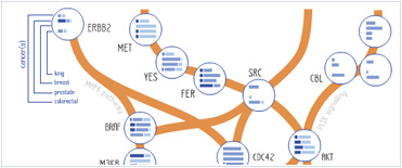


 Foundational characteristics of cancer include proliferation, angiogenesis, migration, evasion of apoptosis, and cellular immortality. Find key markers for these cellular processes and antibodies to detect them.
 The SUMOplot™ Analysis Program predicts and scores sumoylation sites in your protein. SUMOylation is a post-translational modification involved in various cellular processes, such as nuclear-cytosolic transport, transcriptional regulation, apoptosis, protein stability, response to stress, and progression through the cell cycle.
 The Autophagy Receptor Motif Plotter predicts and scores autophagy receptor binding sites in your protein. Identifying proteins connected to this pathway is critical to understanding the role of autophagy in physiological as well as pathological processes such as development, differentiation, neurodegenerative diseases, stress, infection, and cancer.
DVL1 Antibody (Center)
Affinity Purified Rabbit Polyclonal Antibody (Pab)
- SPECIFICATION
- CITATIONS: 1
- PROTOCOLS
- BACKGROUND

Application
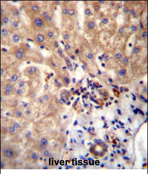
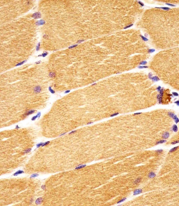
| WB, IF, IHC-P, E |
|---|---|
| Primary Accession | O14640 |
| Other Accession | P54792, Q9WVB9, P51141, NP_004412.2 |
| Reactivity | Human, Mouse |
| Predicted | Rat |
| Host | Rabbit |
| Clonality | Polyclonal |
| Isotype | Rabbit IgG |
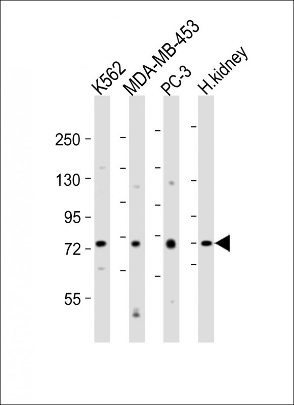
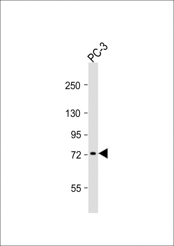
| Calculated MW | 75187 Da |
| Antigen Region | 442-470 aa |
| Gene ID | 1855 |
|---|---|
| Other Names | Segment polarity protein dishevelled homolog DVL-1, Dishevelled-1, DSH homolog 1, DVL1 |
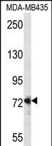
| Target/Specificity | This DVL1 antibody is generated from rabbits immunized with a KLH conjugated synthetic peptide between 442-470 amino acids from the Central region of human DVL1. |
| Dilution | IF~~1:25 WB~~1:1000 IHC-P~~1:25 |
| Format | Purified polyclonal antibody supplied in PBS with 0.09% (W/V) sodium azide. This antibody is purified through a protein A column, followed by peptide affinity purification. |
| Storage | Maintain refrigerated at 2-8°C for up to 2 weeks. For long term storage store at -20°C in small aliquots to prevent freeze-thaw cycles. |
| Precautions | DVL1 Antibody (Center) is for research use only and not for use in diagnostic or therapeutic procedures. |
| Name | DVL1 |
|---|---|
| Function | Participates in Wnt signaling by binding to the cytoplasmic C-terminus of frizzled family members and transducing the Wnt signal to down-stream effectors. Plays a role both in canonical and non-canonical Wnt signaling. Plays a role in the signal transduction pathways mediated by multiple Wnt genes. Required for LEF1 activation upon WNT1 and WNT3A signaling. DVL1 and PAK1 form a ternary complex with MUSK which is important for MUSK-dependent regulation of AChR clustering during the formation of the neuromuscular junction (NMJ). |
| Cellular Location | Cell membrane; Peripheral membrane protein; Cytoplasmic side. Cytoplasm, cytosol. Cytoplasmic vesicle Note=Localizes at the cell membrane upon interaction with frizzled family members. |

Provided below are standard protocols that you may find useful for product applications.
Background
DVL1, the human homolog of the Drosophila dishevelled gene (dsh) encodes a cytoplasmic phosphoprotein that regulates cell proliferation, acting as a transducer molecule for developmental processes, including segmentation and neuroblast specification. DVL1 is a candidate gene for neuroblastomatous transformation. The Schwartz-Jampel syndrome and Charcot-Marie-Tooth disease type 2A have been mapped to the same region as DVL1. The phenotypes of these diseases may be consistent with defects which might be expected from aberrant expression of a DVL gene during development.
References
Metcalfe, C., et al. J. Cell. Sci. 123 (PT 9), 1588-1599 (2010) :
Hu, T., et al. J. Biol. Chem. 285(18):13561-13568(2010)
Varelas, X., et al. Dev. Cell 18(4):579-591(2010)
Jugessur, A., et al. PLoS ONE 5 (7), E11493 (2010) :
Guo, J., et al. PLoS ONE 4 (11), E7982 (2009) :
If you have used an Abcepta product and would like to share how it has performed, please click on the "Submit Review" button and provide the requested information. Our staff will examine and post your review and contact you if needed.
If you have any additional inquiries please email technical services at tech@abcepta.com.
Ordering Information
Other Products
Shipping Information