


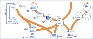


 Foundational characteristics of cancer include proliferation, angiogenesis, migration, evasion of apoptosis, and cellular immortality. Find key markers for these cellular processes and antibodies to detect them.
 The SUMOplot™ Analysis Program predicts and scores sumoylation sites in your protein. SUMOylation is a post-translational modification involved in various cellular processes, such as nuclear-cytosolic transport, transcriptional regulation, apoptosis, protein stability, response to stress, and progression through the cell cycle.
 The Autophagy Receptor Motif Plotter predicts and scores autophagy receptor binding sites in your protein. Identifying proteins connected to this pathway is critical to understanding the role of autophagy in physiological as well as pathological processes such as development, differentiation, neurodegenerative diseases, stress, infection, and cancer.
AP1M1 Antibody (Center)
Affinity Purified Rabbit Polyclonal Antibody (Pab)
- SPECIFICATION
- CITATIONS
- PROTOCOLS
- BACKGROUND

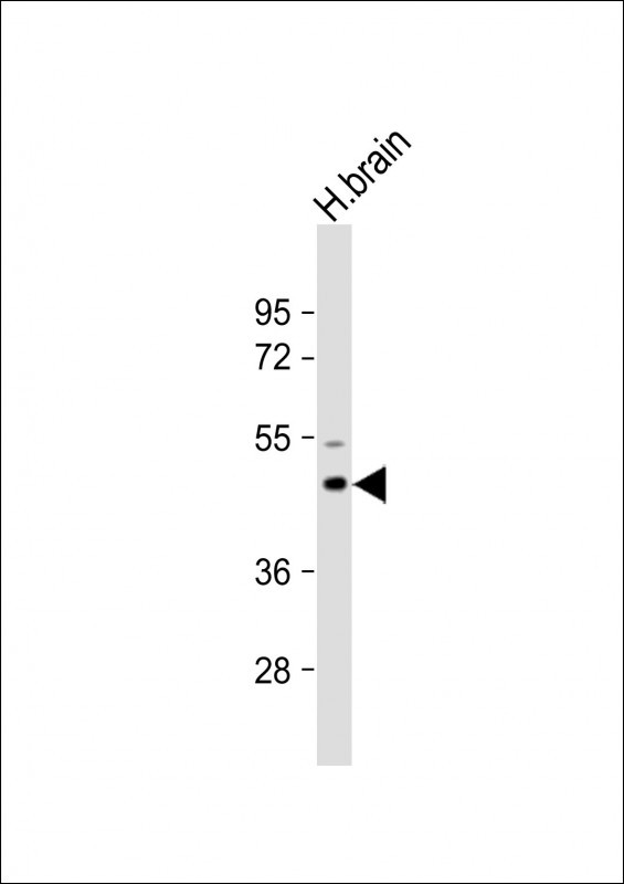
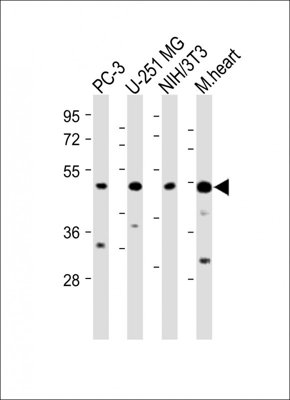
Application
| WB, IF, IHC-P, IHC-P-Leica, E |
|---|---|
| Primary Accession | Q9BXS5 |
| Other Accession | Q32Q06, P35585, Q2KJ81, NP_115882.1 |
| Reactivity | Human, Mouse |
| Predicted | Bovine, Rat |
| Host | Rabbit |
| Clonality | Polyclonal |
| Isotype | Rabbit IgG |
| Calculated MW | 48587 Da |
| Antigen Region | 199-227 aa |
| Gene ID | 8907 |
|---|---|
| Other Names | AP-1 complex subunit mu-1, AP-mu chain family member mu1A, Adaptor protein complex AP-1 subunit mu-1, Adaptor-related protein complex 1 subunit mu-1, Clathrin assembly protein complex 1 mu-1 medium chain 1, Clathrin coat assembly protein AP47, Clathrin coat-associated protein AP47, Golgi adaptor HA1/AP1 adaptin mu-1 subunit, Mu-adaptin 1, Mu1A-adaptin, AP1M1, CLTNM |
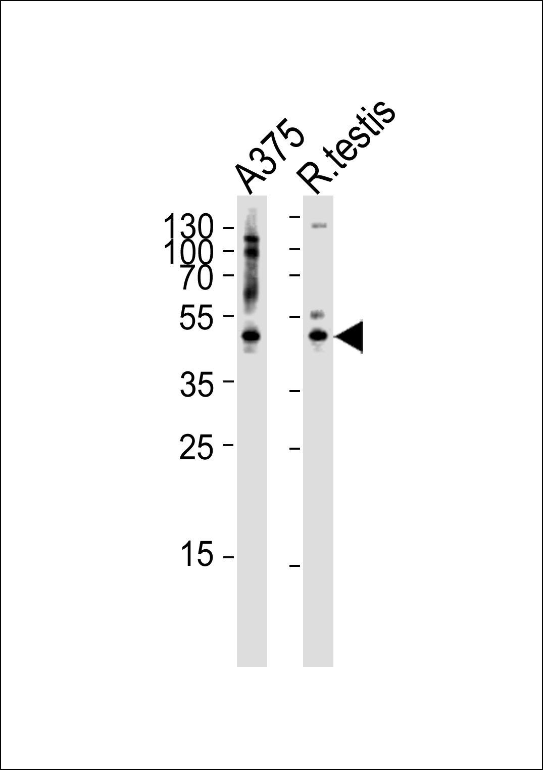
| Target/Specificity | This AP1M1 antibody is generated from rabbits immunized with a KLH conjugated synthetic peptide between 199-227 amino acids from the Central region of human AP1M1. |
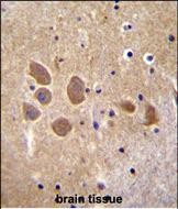
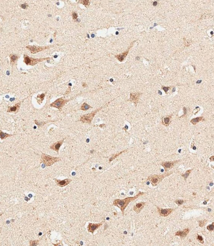
| Dilution | IF~~1:10~50 WB~~1:1000 IHC-P~~1:10~50 IHC-P-Leica~~1:500 |
| Format | Purified polyclonal antibody supplied in PBS with 0.09% (W/V) sodium azide. This antibody is purified through a protein A column, followed by peptide affinity purification. |
| Storage | Maintain refrigerated at 2-8°C for up to 2 weeks. For long term storage store at -20°C in small aliquots to prevent freeze-thaw cycles. |
| Precautions | AP1M1 Antibody (Center) is for research use only and not for use in diagnostic or therapeutic procedures. |
| Name | AP1M1 |
|---|---|
| Synonyms | CLTNM |
| Function | Subunit of clathrin-associated adaptor protein complex 1 that plays a role in protein sorting in the trans-Golgi network (TGN) and endosomes. The AP complexes mediate the recruitment of clathrin to membranes and the recognition of sorting signals within the cytosolic tails of transmembrane cargo molecules. |
| Cellular Location | Golgi apparatus. Cytoplasmic vesicle, clathrin- coated vesicle membrane; Peripheral membrane protein; Cytoplasmic side Note=Component of the coat surrounding the cytoplasmic face of coated vesicles located at the Golgi complex |

Thousands of laboratories across the world have published research that depended on the performance of antibodies from Abcepta to advance their research. Check out links to articles that cite our products in major peer-reviewed journals, organized by research category.
info@abcepta.com, and receive a free "I Love Antibodies" mug.

Provided below are standard protocols that you may find useful for product applications.
Background
The protein encoded by this gene is the medium chain of the trans-Golgi network clathrin-associated protein complex AP-1. The other components of this complex are beta-prime-adaptin, gamma-adaptin, and the small chain AP1S1. This complex is located at the Golgi vesicle and links clathrin to receptors in coated vesicles. These vesicles are involved in endocytosis and Golgi processing. Alternatively spliced transcript variants encoding distinct protein isoforms have been found for this gene. [provided by RefSeq].
References
Sawasdee, N., et al. Biochem. Biophys. Res. Commun. 401(1):85-91(2010)
Venkatesan, K., et al. Nat. Methods 6(1):83-90(2009)
Noviello, C.M., et al. J. Virol. 82(3):1249-1258(2008)
Medigeshi, G.R., et al. Traffic 9(1):121-132(2008)
Roeth, J.F., et al. J. Cell Biol. 167(5):903-913(2004)
If you have used an Abcepta product and would like to share how it has performed, please click on the "Submit Review" button and provide the requested information. Our staff will examine and post your review and contact you if needed.
If you have any additional inquiries please email technical services at tech@abcepta.com.
Ordering Information
Other Products
Shipping Information